SEARCH:

Dental Management of Asthmatic Patient

Dental management of Medically compromised patient _ Asthma
Dental Management of Asthmatic Patient

Asthma is a respiratory disease that causes reversible airway obstruction and a reduced ability to expire or completely empty the lungs of gases. Inflammation is a component of the disease process and results in increased mucous secretions in the lungs and swelling in the bronchioles.

Clinical Manifestations include:

1. Cough
2. Shortness of breath
3. Chest tightness
4. Wheezing.
5. Increased heart rate
6. Nervousness
7. Sweating

The most common form, called extrinsic asthma, develops as a result of allergy to environmental pollutants. It generally occurs during childhood and may or may not extend into adult years.

A second type of asthma is intrinsic asthma, or infectious asthma, is A non-allergic form of asthma usually first occurring later in life that tends to be chronic and persistent rather than episodic , most often seen in patients older than age 35. Unlike the extrinsic asthma patient, this patient may exhibit a chronic cough with sputum production between attacks. Intrinsic asthma usually occurs as a result of some type of bronchial infection.

Oral Complications associated with asthma medications include dry mouth, candidiasis, and an increased dental caries rate.

TREATMENT (Dental Management of Asthmatic Patient)
1. Stop all dental treatment. Be sure to remove all materials and instruments from the patient’s mouth.


2. Position the patient. Raise the patient upright; since the patient will be struggling for air, it will be easier for the patient to breathe if seated upright.


3. Use a bronchodilator. The patient’s bronchodilator should be placed within easy reach in case an attack occurs. If an attack does take place, allow the patient to administer the bronchodilator; patients know what their usual dose involves . The bronchodilator is an aerosol medication that usually includes epinephrine, which relaxes the bronchioles and makes it easier for the patient to breathe.


4. Administer oxygen.


5. Administer epinephrine or another drug intravenously it may be necessary, if the bronchodilator does not relieve the attack.

MCQs on Instruments Used in Oral Surgery

# A straight elevator is properly used to advantage when the:
A. Adjacent tooth is the fulcrum
B. Tooth is isolated
C. Interdental bone is fulcrum
D. Adjacent tooth is not to be extracted

# Most of the elevators used in exodontias works on the principle
A. Class I lever
B. Class II lever
C. Class III lever
D. Wheel and axle

MCQs on Skin and vesiculobullous Lesions - Oral Medicine MCQs - Oral Pathology MCQs

 # All of the following lesions may be classified as Odontogenic Tumors EXCEPT
A. Acanthomatous ameloblastoma
B. Branchial cleft cyst
C. Myxoma
D. Simple ameloblastoma

# Fish net pattern is pemphigus vulgaris is seen in which of the following tests?
A. Direct immunofluorescence
B. Tzanck smear
C. FNAC
D. Histopathology

# All of the following are inherited disorders of connective tissue EXCEPT:
A. Alport syndrome
B. Ehlers-Danlos syndrome
C. Marfan syndrome
D. McArdle’s disease

MCQs on Oral Mucous Membrane - Oral Anatomy and Histology

 # Which of the following is correct:
A. Non-keratinized epithelium is characterized by absence of stratum granulosum and stratum corneum, The surface cells are nucleated
B. Para-keratinized epithelium is characterized by superficial cells with pyknotic nuclei and absence of stratum granulosum
C. In ortho keratinization the superficial cells lose their nuclei, but stratum granulosum is present
D. All of the above

# Long connective tissue papillae and keratinized epithelia are a feature of these parts of oral mucosa:
A. gingiva and alveolar mucosa
B. Hard palate and gingiva
C. Buccal and alveolar mucosa
D. Hard and soft palate

# All of the following is lined by stratified squamous epithelium, except:
A. Lips
B. Tongue
C. Roof of the soft palate
D. Oropharynx

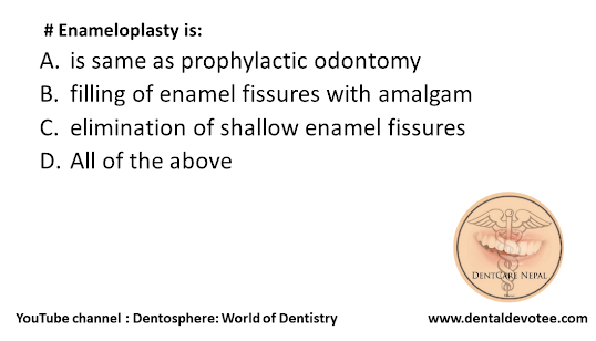
Enameloplasty is:

  # Enameloplasty is:
A. is same as prophylactic odontomy
B. filling of enamel fissures with amalgam
C. elimination of shallow enamel fissures
D. All of the above


The correct answer is C. Elimination of shallow enamel fissures.

Historically, enameloplasty was utilized as an ultraconservative procedure on the occlusal surfaces, which were deemed to be at risk of the development of a pit or issure caries lesion. Extreme prudence was exercised in the selection of these areas and in the depth of enamel removed. This procedure was never used unless the area could be transformed into a cleansable groove (or fossa) by a minimal reduction of enamel, and unless occlusal contacts could be maintained. This procedure technically included a preparation stage but no restoration stage. Currently, clinical situations such as these (ICDAS 1 or 2) are managed by treatment with luoride or placement of sealants. Research studies support the filling of issures/pits and narrow grooves/fossae (i.e., “sealing”) with low viscosity composite resin materials, without any mechanical alteration (enameloplasty) of the at-risk tooth anatomy.

Additionally, “prophylactic odontotomy” procedures were used in the past. These more aggressive procedures involved preparing developmental or structural imperfections of the enamel that were thought to be at increased risk of caries and filling the preparation with amalgam to prevent caries from developing in these sites. Prophylactic odontotomy is no longer advocated as a preventive measure.

Reference: Sturdevant’s Art and Science of Operative Dentistry, Seventh Edition, Page No 125

# Spontaneous bleeding usually occurs with a platelet count of:

 # Spontaneous bleeding usually occurs with a platelet count of:
A. Less than 50000/mm3
B. 50000-75000/ mm3
C. 75000-100000/ mm3
D. 100000-150000/mm3


The correct answer is: A. Less than 50000/mm3.

Spontaneous bleeding does not usually occur until the platelet count falls below 20 × 109/L, unless their
function is also compromised. Purpura and spontaneous bruising are characteristic but there may also be oral, nasal, gastrointestinal or genitourinary bleeding. Severe thrombocytopenia (< 10 × 109/L) may result in retinal haemorrhage and potentially fatal intracranial bleeding, but this is rare.

Reference: Davidson's Principles and Practice of Medicine, 22nd Edition, Page No. 1008.

Chancre most commonly occurs on:

 # Chancre most commonly occurs on:
A. Genitals
B. Tonsils
C. Tongue
D. Floor of the mouth


The correct answer is A. Genitals.

A chancre is a painless genital ulcer most commonly formed during the primary stage of syphilis. This infectious lesion forms approximately 21 days after the initial exposure to Treponema pallidum, the gram-negative spirochaete bacterium yielding syphilis. Chancres transmit the sexually transmissible disease of syphilis through direct physical contact. These ulcers usually form on or around the anus, mouth, penis and vagina. Chancres may diminish between four and eight weeks without the application of medication.

Chancres, as well as being painless ulcerations formed during the primary stage of syphilis, are associated with the African trypanosomiasis sleeping sickness, surrounding the area of the tsetse fly bite.