SEARCH:

Saw toothed Rete Pegs are seen in:

 #  Saw toothed Rete Pegs are seen in:
A. Lichen planus
B. Lichenoid reaction
C. Leukoplakia
D. Leukoedema


The correct answer is A. Lichen Planus.

Histopathologic examination of lesional tissue is the most relevant investigation in cases of Oral Lichen Planus. Typical findings include hyperparakeratosis or hyperorthokeratosis with thickening of the granular layer, acanthosis with intracellular edema of the spinous cells in some instances, the development of a ‘saw tooth’ appearance of the rete pegs. Band-like subepithelial mononuclear infiltrate consisting of T-cells and histiocytes; increased numbers of intraepithelial T-cells; and degenerating basal keratinocytes that form colloid (Civatte, hyaline, cytoid) bodies, which appear as homogenous eosinophilic globules are consistently seen.


Bleeding joints is a characteristic feature of:

 #  Bleeding joints is a characteristic feature of:
A. Vitamin C deficiency
B. Vitamin K deficiency
C. Hemophilia
D. Thrombocytopenia


The correct answer is C. Hemophilia.

Patients with hemophilia exhibit persistent bleeding, either spontaneous or following even slight trauma that produces the mildest of abrasions or cuts. Hemorrhage into the subcutaneous tissues, internal organs, and joints is also a common feature and may result in massive hematomas.

An asymptomatic tooth has deep caries on occlusal surface. Radiograph shows radiopaque mass at apex of the tooth. This mass is most likely to be:

 # An asymptomatic tooth has deep caries on occlusal surface. Radiograph shows radiopaque mass at apex of the tooth. This  mass is most likely to be:
A. Cementoma
B. Condensing osteitis
C. Chronic apical periodontitis
D. Acute apical periodontitis


The correct answer is B. Condensing osteitis.

In condensing osteitis, radiographs demonstrate radiopacity which is not attached to tooth, entire root outline is visible, lamina dura intact and periodontal ligament is widened. These features differentiate it from cementoblastoma, in which the radiopacity is attached to tooth, lamina dura absent and root outline is not visible. In condensing osteitis the tooth may be vital or non-vital.

In a leukemic patient, bleeding does not stop because:

 # In a leukemic patient, bleeding does not stop because:
A. Decreased platelet count 
B. Increased WBC count
C. Decreased calcium level
D. Decrease in coagulation level


The correct answer is A. Decreased Platelet Count

The sign and symptoms of acute leukemia result from either bone marrow suppression or infiltration
of leukemic cells into organs tissues. These changes cause anemia, thrombocytopenia and a decrease in
neutrophils. Thrombocytopenia causes spontaneous bleeding such as petechiae, ecchymoses, epistaxis,
melena. It may necessary to perform surgery on patients with platelet counts in the range of 25,000
because of the difficulty in achieving platelet levels due to circulating platelet antibodies.


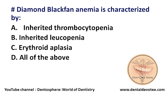
Diamond Blackfan anemia is characterized by:

 # Diamond Blackfan anemia is characterized by:
A. Inherited thrombocytopenia 
B. Inherited leucopenia
C. Erythroid aplasia
D. All of the above


The correct answer is C. Erythroid aplasia.

Diamond–Blackfan anemia (DBA) is a congenital erythroid aplasia that usually presents in infancy. DBA causes low red blood cell counts (anemia), without substantially affecting the other blood components (the platelets and the white blood cells), which are usually normal. This is in contrast to Shwachman–Bodian–Diamond syndrome, in which the bone marrow defect results primarily in neutropenia, and Fanconi anemia, where all cell lines are affected resulting in pancytopenia.

First noted by Hugh W. Josephs in 1936, the condition is however named for the pediatricians Louis K. Diamond and Kenneth Blackfan, who described congenital hypoplastic anemia in 1938. 


Reduction of flow of saliva is not generally seen in:

# Reduction of flow of saliva is not generally seen in:
A. Elderly diabetes
B. Patient undergoing radiation
C. Patient suffering from parkinsonism
D. Patient of Phenothiazine drugs


The correct answer is C. Patient suffering from Parkinsonism.

Xerostomia : It is the subjective clinical condition of less than normal amount of saliva. It is dryness of mouth, which is a clinical manifestation of salivary gland dysfunction.

Causes:
- Radiation induced
- Pharmacologically induced xerostomia—there are about 500 drugs which can cause xerostomia. The classes of drugs which cause xerostomia include anticonvulsants, antiemetics, antihistaminics, anti-hypertensives and antispasmodics. The mode of action for decreased salivary flow is generally related to the para-sympathetic activity, usually an antimuscarine effect.
- Smoking, Mouth breathing
- Developmental—developmental abnormalities of salivary glands, tumors, autoimmune states and certain diseases which affect afferent or efferent portions of neural transmission reflex
- Systemic alternations resulting in xerostomia 
• Nutritional—certain deficiency states like pernicious anemia, iron deficiency anemia and deficiency of
vitamin A and hormones can cause xerostomia.
• Fluid loss—fluid loss associated with hemorrhage, sweating, diarrhea, vomiting.
• Diabetes mellitus—it is associated with xerostomia.
• Sjögren syndrome—xerostomia is also common in Sjögren syndrome.
• Other disease—systemic diseases, which are accompanied by high temperature and dehydration, usually result in diminished salivation. Xerostomia may also be found in HIV infection, sarcoidosis, and graft versus host resistance.

Marked reduction in the amount of dentin, widening of predentin layer, presence of large areas of interglobular dentin and irregular pattern of dentin

# Which of the following condition shows marked reduction in the amount of dentin, widening of predentin layer, presence of large areas of interglobular dentin and irregular pattern of dentin?
A. Hypocalcified dentin
B. Regional odontodysplasia
C. Dentin dysplasia
D. Dentinogenesis imperfecta


The correct answer is B. Regional odontodysplasia.

The most characteristic features of Regional odontodysplasia are the marked reduction in the amount of dentin, the widening of the predentin layer, the presence of large areas of interglobular dentin, and an irregular tubular pattern of dentin.