SEARCH:

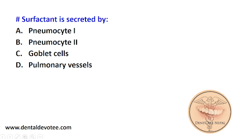
Surfactant is secreted by:

 # Surfactant is secreted by:
A. Pneumocyte I
B. Pneumocyte II
C. Goblet cells
D. Pulmonary vessels


The correct answer is B. Pneumocyte II.

Surfactant lowers the surface tension of the fluid lining of the alveoli and prevents the collapsing tendency of the lungs. It is a phospholipid. The absence of surfactant results in Hyaline membrane disease (or) respiratory distress syndrome. 

Scleroderma is associated with:

 # Scleroderma is associated with:
A. Parry Romberg Syndrome
B. Pierre Robin Syndrome
C. Gardner Syndrome
D. Peutz Jegher Syndrome


The correct answer is A. Parry Romberg Syndrome.

Hemifacial atrophy or Parry Romberg syndrome consists of slow progressive atrophy of soft tissues of essentially half the face, characterized by progressive wasting of subcutaneous fat, accompanied by atrophy of skin, cartilage, bone, and muscle. This syndrome is accompanied usually by contralateral Jacksonian epilepsy, trigeminal neuralgia, and changes in hair and eyes. It is also reported that hemifacial atrophy is a form of localized scleroderma which is supported by its concurrence with scleroderma. 


Samaya Dental Care Pvt.Ltd., Kadaghari Kathmandu

 Name of Dental Clinic: Samaya Dental Care Pvt.Ltd.
Address (Full): Medical chowk, Kadaghari-9,Kathmandu
Year of Establishment: 2079 B. S.
Name of the chief Dental Surgeon: Dr.Nisha Sapkota
CONTACT NUMBER: 014492979
NMC Number of Dental Surgeon: 29031

Some Sample Intraoral Photograph in Orthodontics

 Intraoral photography involves capturing a series of detailed images of the inside of a patient's mouth to document their oral health, aid in diagnosis, and monitor treatment progress. To provide a comprehensive view of the patient's oral structures, several different views or types of intraoral photographs are commonly taken. These views offer orthodontists and other dental professionals a well-rounded understanding of the patient's dental condition. Here are some of the most common views included in intraoral photography: 

Frontal View (Full-Arch View): This view captures the entire arch of teeth from the front, providing a comprehensive overview of tooth alignment, spacing, and symmetry. It helps orthodontists assess the overall aesthetics of the smile and identify any irregularities. 

Right Lateral View: The right lateral view is taken from the side to evaluate the profile of the teeth and jaws from the right angle. This view is essential for diagnosing issues like overbites, underbites, and jaw discrepancies.


Left Lateral View: Similar to the right lateral view, the left lateral view provides a profile perspective from the left side. It aids in assessing the relationship between the upper and lower jaws and identifying any asymmetries.

Upper Occlusal View: The upper occlusal view captures the biting surfaces of the upper teeth when the patient bites down. It helps orthodontists evaluate the alignment of upper teeth, spacing, and the overall occlusal relationship.

Lower Occlusal View: Similar to the upper occlusal view, the lower occlusal view focuses on the biting surfaces of the lower teeth. This view assists in evaluating lower arch alignment, spacing, and occlusal considerations.

These five specific views are crucial for orthodontists to accurately diagnose dental conditions, plan appropriate treatments, and monitor progress throughout the orthodontic journey.


Intraoral Photography in Orthodontics

 Orthodontics, the field of dentistry dedicated to correcting misaligned teeth and jaws, has evolved significantly over the years. With the advent of cutting-edge technologies, orthodontists now have a vast array of tools at their disposal to provide more accurate diagnoses, personalized treatment plans, and improved patient experiences. Among these tools, intraoral photography stands out as a valuable technique that has revolutionized the way orthodontic professionals document, analyze, and communicate treatment progress.

The Power of Intraoral Photography

Intraoral photography involves capturing high-quality images of the inside of a patient's mouth, allowing orthodontists to closely examine teeth, gums, and oral structures in intricate detail. This technique offers a multitude of advantages that contribute to more effective orthodontic treatments and enhanced patient satisfaction.

1. Precise Documentation:
Gone are the days of relying solely on hand-drawn diagrams or vague written descriptions to document a patient's oral condition. Intraoral photography provides orthodontists with clear, high-resolution images that accurately record a patient's initial condition and subsequent progress. These images can be easily stored in digital patient files and referred to at any stage of treatment, aiding in comprehensive case analysis and treatment evaluation.

2. Visual Communication:
One of the challenges in orthodontics is effectively communicating treatment plans and progress to patients. Intraoral photographs allow orthodontists to show patients visual evidence of their oral health status, making it easier for them to understand the issues that need to be addressed. Patients can see exactly what the orthodontist sees, fostering a sense of trust and understanding between the patient and the healthcare provider.

3. Treatment Planning and Monitoring:
Intraoral photographs offer orthodontists a unique perspective into a patient's oral structures that might not be as apparent during a routine clinical examination. These detailed images aid in the accurate diagnosis of various dental conditions and help orthodontists devise precise treatment plans tailored to each patient's needs. Moreover, the ability to capture images at different stages of treatment enables orthodontists to monitor progress closely and make any necessary adjustments to ensure the desired outcomes are achieved.


4. Education and Informed Consent:
When patients are well-informed about their treatment options and the expected outcomes, they are more likely to comply with treatment plans and experience greater satisfaction. Intraoral photographs allow orthodontists to visually explain different treatment modalities, potential challenges, and expected results. This informed consent process empowers patients to actively participate in their treatment journey, leading to more positive treatment experiences.

5. Research and Collaboration:
Intraoral photography not only benefits individual patients but also contributes to the advancement of orthodontic research. The ability to capture standardized, high-quality images aids in data collection and analysis for research studies aimed at improving orthodontic techniques and outcomes. Additionally, these images can be shared efficiently among colleagues and specialists, promoting collaborative discussions and multidisciplinary approaches to complex cases.

Incorporating Intraoral Photography into Practice

Implementing intraoral photography into orthodontic practice requires specialized equipment, including intraoral cameras capable of capturing detailed images of the oral cavity. These cameras are designed to be comfortable for patients and easy for orthodontic professionals to use. The images captured by these cameras can then be integrated into digital patient records and treatment planning software for seamless organization and analysis.

Orthodontists and their teams can benefit from training in proper intraoral photography techniques to ensure consistent, high-quality image capture. Learning how to position the camera, control lighting conditions, and select appropriate settings are all essential skills for obtaining accurate images that provide meaningful diagnostic and treatment information.

Conclusion

Intraoral photography has emerged as a powerful tool in orthodontics, enhancing how orthodontic professionals diagnose, plan, and monitor treatments. By providing detailed visual records, facilitating patient communication, aiding in treatment planning, and contributing to research endeavors, intraoral photography has become an indispensable aspect of modern orthodontic practice. As technology continues to advance, intraoral photography will likely continue to evolve, further improving the precision and efficacy of orthodontic treatments for patients of all ages.

Determining the Need for Dental Scaling: A Guide for Home Assessment

 Maintaining optimal oral hygiene is vital for overall health and well-being. One crucial aspect of oral care is dental scaling, a professional procedure aimed at removing stubborn plaque and tartar buildup from your teeth and gums. While it's always best to consult a dentist for a proper assessment, there are several indicators you can consider to gauge whether your teeth might need scaling. Here's a guide to help you assess the need for dental scaling at home:

Understanding Dental Scaling: Dental scaling, also known as deep cleaning, is a preventive dental procedure designed to remove accumulated plaque and tartar that regular brushing and flossing can't eliminate. Plaque is a sticky film of bacteria that adheres to your teeth and, if not removed, can lead to tooth decay, gum disease, and other oral health issues. Over time, plaque can harden into tartar, which cannot be removed through regular brushing and requires professional intervention.

Signs That Your Teeth Might Need Scaling: While self-assessment can provide some insight, it's essential to remember that only a qualified dentist can provide a definitive diagnosis. However, here are some signs that might suggest the need for dental scaling:

             1. Visible Tartar Buildup: Gently examine your teeth in a well-lit area using a mirror. If you notice a yellowish or brownish substance adhering to the tooth surfaces, especially near the gumline, as shown in the picture below, this could be tartar buildup.


              2. Bleeding Gums: If your gums bleed while brushing, flossing, or eating, it might be a sign of gum inflammation, commonly known as gingivitis. While bleeding gums can result from various factors, it can be an indication that plaque and tartar are irritating your gums.

             3. Persistent Bad Breath: Foul breath that doesn't improve with regular brushing, flossing, and mouthwash might be an indicator of bacteria buildup in hard-to-reach areas of your mouth. These bacteria could be residing in plaque and tartar.

              4. Receding Gums: If you notice that your gums are pulling away from your teeth, making your teeth appear longer, it could be a sign of advanced gum disease. Scaling might be necessary to remove tartar below the gumline and prevent further damage.

               5. Sensitivity: Increased tooth sensitivity to hot, cold, sweet, or acidic foods and beverages might suggest gum recession due to tartar accumulation. The exposure of sensitive tooth surfaces can lead to discomfort.

               6. Visible Cavities: If you can see or feel small holes or pits in your teeth, these could be cavities. Dental scaling might be needed to remove the plaque and tartar surrounding the cavities before they worsen.


Steps to Maintain Good Oral Hygiene:

While self-assessment can be helpful, it's important to consult a dentist for a professional evaluation and treatment recommendations. In the meantime, follow these steps to maintain excellent oral hygiene:

- Brush Regularly: Brush your teeth at least twice a day with a fluoride toothpaste to remove plaque and prevent tartar buildup.

- Floss Daily: Flossing helps remove food particles and plaque from between your teeth and along the gumline.

- Use Mouthwash: An antimicrobial mouthwash can help reduce bacteria in your mouth, promoting fresher breath and healthier gums.

- Maintain a Balanced Diet: Consume a diet rich in fruits, vegetables, lean proteins, and whole grains. Limit sugary and acidic foods and beverages that can contribute to tooth decay.

- Stay Hydrated: Drinking water helps rinse away food particles and bacteria from your mouth, reducing the risk of plaque buildup.

- Regular Dental Checkups: Visit your dentist for regular checkups and cleanings every six months. Professional cleanings can prevent tartar accumulation and catch dental issues early.

Final Thoughts:

While these self-assessment methods can give you some indication of your oral health status, they are not a substitute for professional dental care. If you notice any signs of plaque, tartar buildup, gum inflammation, or other oral health concerns, it's crucial to schedule an appointment with a dentist. Dental scaling, if recommended, can prevent more severe oral health issues and contribute to your overall well-being. Remember, proactive oral care is the key to a healthy smile.

For best result in class III malocclusion correction the orthopedic appliance should be worn at least:

 # For best result in class III malocclusion correction the orthopedic appliance should be worn at least:
A. 12-16 hours per day
B. 8-10 hours per day
C. 5-8 hours per day
D. Throughout night


The correct answer is A. 12-16 hours per day.

Chin cup and  face mask are the orthopedic appliances that can be given to correct skeletal class III malocclusion. At least 12 to 14 hours of wear per day produces desired results.